
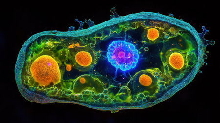
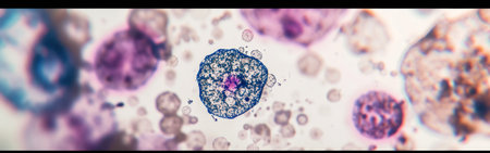
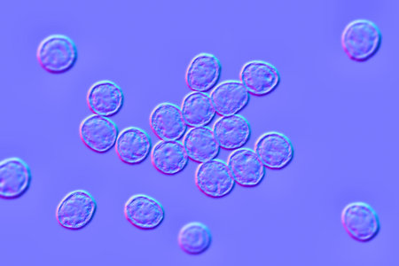
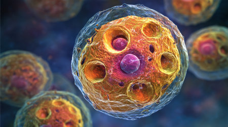
A highly detailed 3D illustration showing various bacteria and microbes in different colors, providing a close-up microscopic view of microorganisms
Коллекция по умолчанию
Коллекция по умолчанию
Создать новую
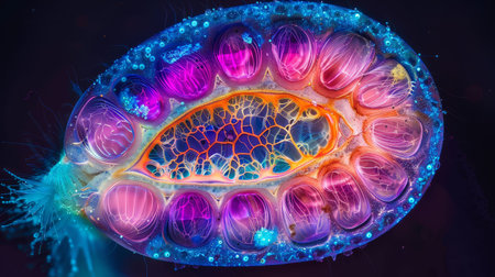
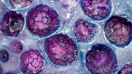
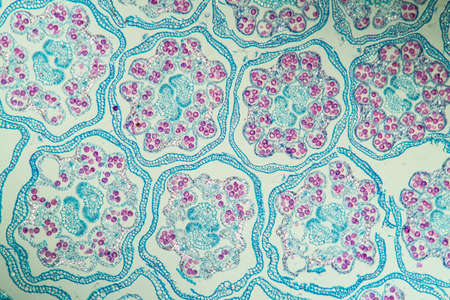
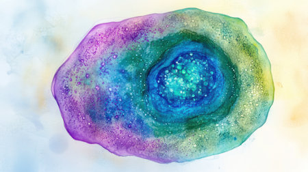
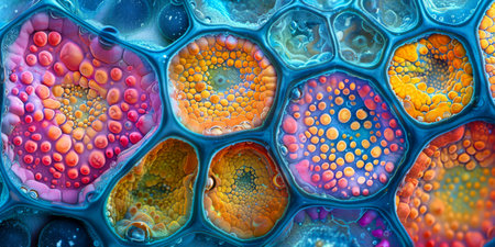
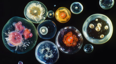
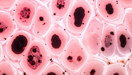
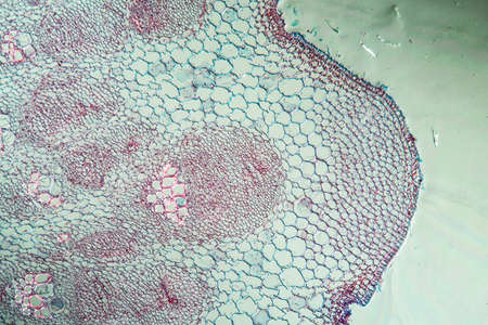
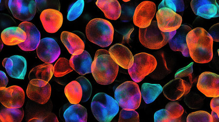
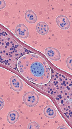
Vibrant cross-section of a developing seed under UV light, highlighting the embryo and endosperm in bright colors
Коллекция по умолчанию
Коллекция по умолчанию
Создать новую
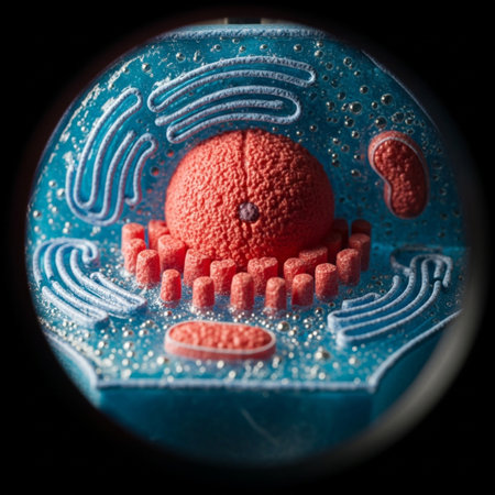
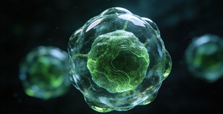
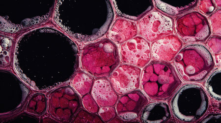
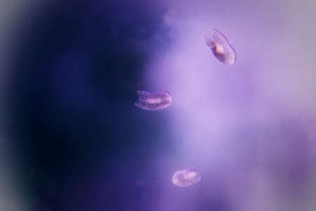
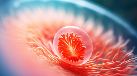
A tightly framed, high-definition close-up (hero detail) of the central nucleus of a simplified, red
Коллекция по умолчанию
Коллекция по умолчанию
Создать новую

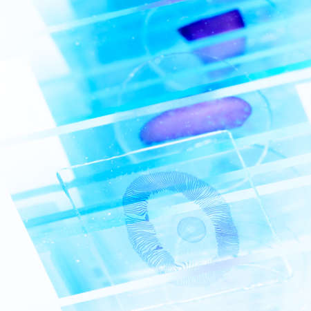
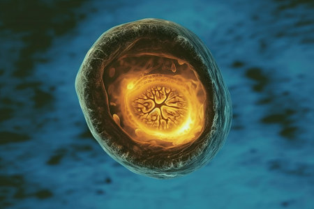
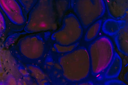
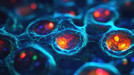
Rows of microscope glass slide in the cells
Коллекция по умолчанию
Коллекция по умолчанию
Создать новую

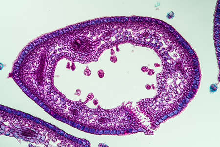
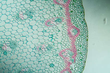
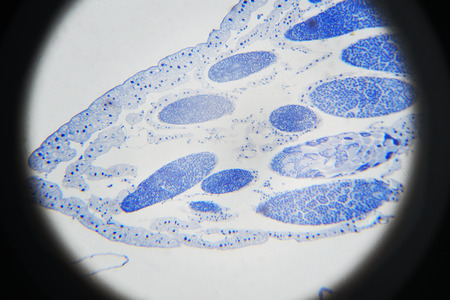
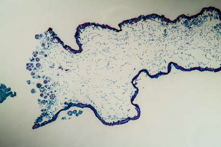
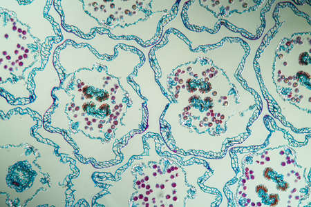
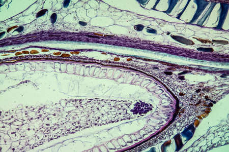
Cross-section leaf Plant of under the microscope for classroom education.
Коллекция по умолчанию
Коллекция по умолчанию
Создать новую
science medical glass microscope slide with sample
Коллекция по умолчанию
Коллекция по умолчанию
Создать новую
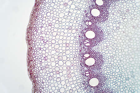
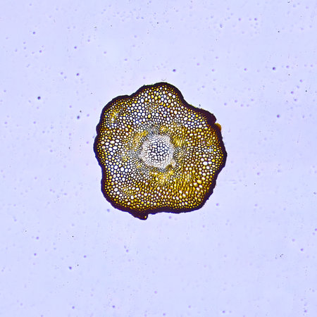
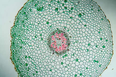
Root plant show root vascular tissue under light microscope view for education.
Коллекция по умолчанию
Коллекция по умолчанию
Создать новую
Microscopic view of the bone marrow stroma shows colonies of mesenchymal stem cells which have the ability to differentiate into various cell types
Коллекция по умолчанию
Коллекция по умолчанию
Создать новую
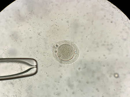
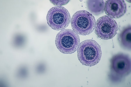
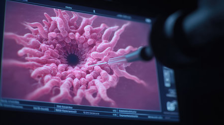
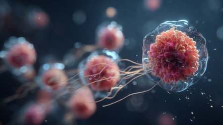
View through microscope at in vitro fertilization process
Коллекция по умолчанию
Коллекция по умолчанию
Создать новую
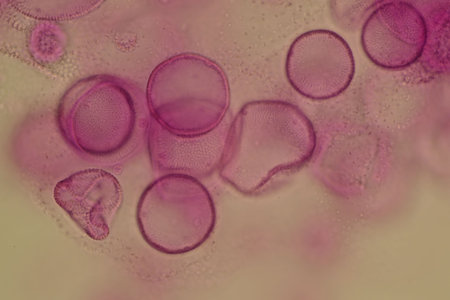
Heather leaf cross section under the microscope, 200x
Коллекция по умолчанию
Коллекция по умолчанию
Создать новую
Close-up view of colorful cellular structures, highlighting unique patterns and textures, illustrating the complexity of microscopic life and its natural beauty
Коллекция по умолчанию
Коллекция по умолчанию
Создать новую
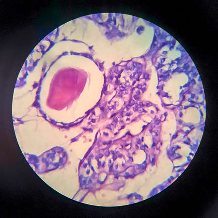
Pancreas cancer, light micrograph, photo under microscope
Коллекция по умолчанию
Коллекция по умолчанию
Создать новую

Microscopic Fungi samples over white background
Коллекция по умолчанию
Коллекция по умолчанию
Создать новую
Histopathology of human kidney cross section under microscope for education.
Коллекция по умолчанию
Коллекция по умолчанию
Создать новую

Rows of microscope glass slide in the cells
Коллекция по умолчанию
Коллекция по умолчанию
Создать новую
A close-up of healthy cells magnified under a light microscope. The cells exhibit detailed structures and activities, showing the complexity of life at a microscopic level.
Коллекция по умолчанию
Коллекция по умолчанию
Создать новую

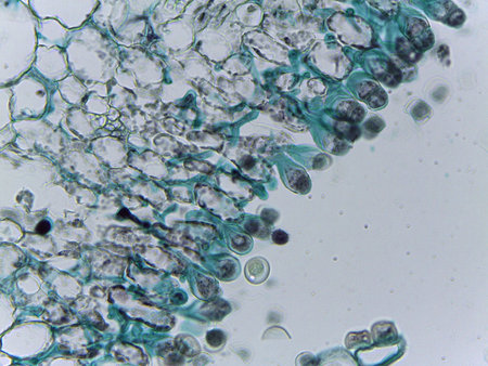
Host cells with spores (mold) are inside wood under the microscope for education.
Коллекция по умолчанию
Коллекция по умолчанию
Создать новую
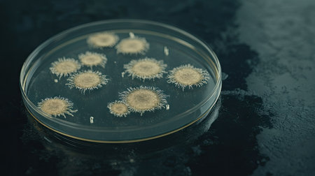
Close-up of a single Petri dish with visible microorganism colonies, on a reflective lab surface, representing research in microbiology.
Коллекция по умолчанию
Коллекция по умолчанию
Создать новую
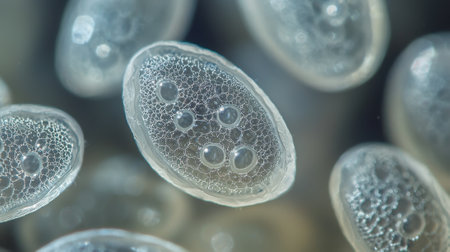
A close up of many small, round, clear objects with a lot of holes in them. The objects are all different sizes and are scattered throughout the image. Scene is one of curiosity and wonder
Коллекция по умолчанию
Коллекция по умолчанию
Создать новую
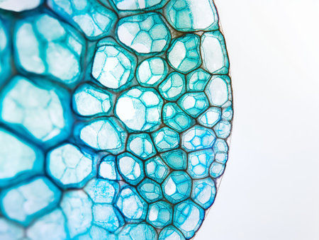
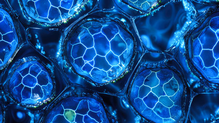
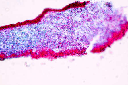
Macro view of a blue stained cellular structure with natural patterns.
Коллекция по умолчанию
Коллекция по умолчанию
Создать новую
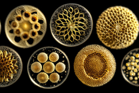
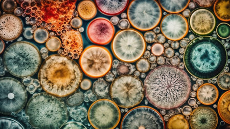
Intricate golden-hued circular patterns resembling microscopic organisms, each with unique designs, highlighted against a contrasting background, showing delicate textures and symmetry
Коллекция по умолчанию
Коллекция по умолчанию
Создать новую
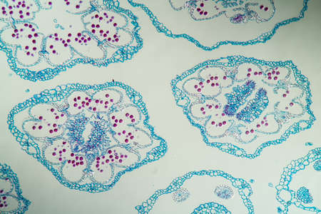
Tansy inflorescence in cross section 100x
Коллекция по умолчанию
Коллекция по умолчанию
Создать новую
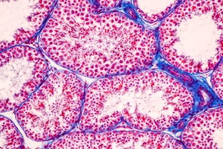
Anatomy and Histological Epididymis and Testis human cells under microscope.
Коллекция по умолчанию
Коллекция по умолчанию
Создать новую
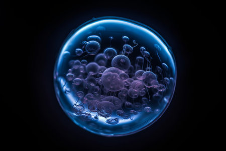
A celestial orb of electric blue hue surrounded by clusters of purple bubbles suspended in a liquidlike atmosphere against a dark backdrop
Коллекция по умолчанию
Коллекция по умолчанию
Создать новую
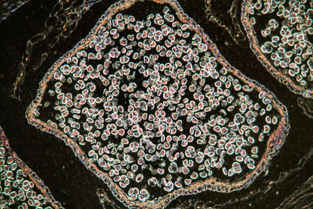
Club moss flower with seeds under the microscope 100x
Коллекция по умолчанию
Коллекция по умолчанию
Создать новую
Microbiology loop isolated on white background
Коллекция по умолчанию
Коллекция по умолчанию
Создать новую
Tansy inflorescence in cross section 100x
Коллекция по умолчанию
Коллекция по умолчанию
Создать новую
Rust fungus on milkweed plant, 200x
Коллекция по умолчанию
Коллекция по умолчанию
Создать новую
Microscopic View of Plant Cell Structures
Коллекция по умолчанию
Коллекция по умолчанию
Создать новую
microscope slides of blood samples indicating rare diseases, created with generative ai
Коллекция по умолчанию
Коллекция по умолчанию
Создать новую
amazing inhabitants of the microworld under a microscope
Коллекция по умолчанию
Коллекция по умолчанию
Создать новую

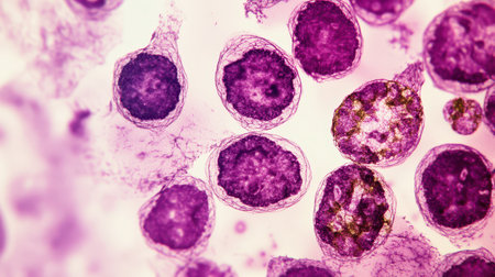
blood cells and expression of mitosis and meiosis of cells
Коллекция по умолчанию
Коллекция по умолчанию
Создать новую
Turmeric with stem in cross section 100x
Коллекция по умолчанию
Коллекция по умолчанию
Создать новую
Microscopic view of blood cells. Microscopic view of blood cells.
Коллекция по умолчанию
Коллекция по умолчанию
Создать новую
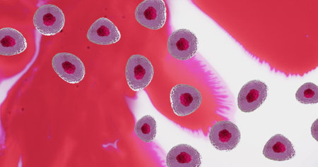
Image of micro of red and pink cells over red and white background. Global science, research and medicine concept digitally generated image.
Коллекция по умолчанию
Коллекция по умолчанию
Создать новую
This cross-section shows a plant cell's structure, featuring green chloroplasts, orange mitochondria, a translucent vacuole, and a striking blue nucleus at the center.
Коллекция по умолчанию
Коллекция по умолчанию
Создать новую
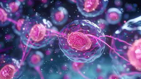
A detailed artwork showcases a single cell featuring a softly glowing blue nucleus and shimmering green and purple cytoplasm. The background gradient enhances its vivid colors and intricate details.
Коллекция по умолчанию
Коллекция по умолчанию
Создать новую
3D illustration of a microscopic close-up of a virus cells
Коллекция по умолчанию
Коллекция по умолчанию
Создать новую
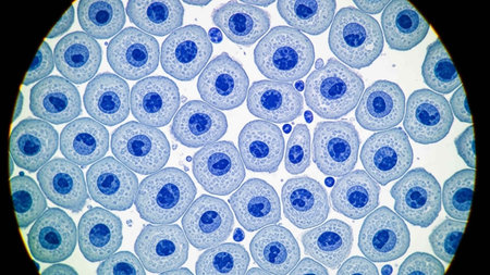
Microscopic photography. Grasshopper meiosis.
Коллекция по умолчанию
Коллекция по умолчанию
Создать новую

Epithelial cells with bacteria from the oral cavity 200x
Коллекция по умолчанию
Коллекция по умолчанию
Создать новую

Abstract science background- pyloric division of the stomach of the dog. Cell biology
Коллекция по умолчанию
Коллекция по умолчанию
Создать новую
Thyroid follicular carcinoma, light micrograph, photo under microscope
Коллекция по умолчанию
Коллекция по умолчанию
Создать новую

Lesser celandine with roots 100x across
Коллекция по умолчанию
Коллекция по умолчанию
Создать новую
This close-up image depicts cancer cells under laboratory equipment, showcasing intricate details for medical analysis and health research. Ideal for educational use.
Коллекция по умолчанию
Коллекция по умолчанию
Создать новую
Vibrant Microscopic Cells Close-Up - Abstract Biological Background.
Коллекция по умолчанию
Коллекция по умолчанию
Создать новую

Bracken stems in cross section 100x
Коллекция по умолчанию
Коллекция по умолчанию
Создать новую
Abstract background with blue and purple watercolor bubbles on a light sky-blue backdrop. --chaos 30 --ar 16:9 --v 6.1 Job ID: f22f57a0-c1f2-4296-9a5d-e73429531fd7
Коллекция по умолчанию
Коллекция по умолчанию
Создать новую

Stomach tissue under the microscope 100x
Коллекция по умолчанию
Коллекция по умолчанию
Создать новую

Living healthy cells (mitosis) - original micro-photo of tissue under a microscope
Коллекция по умолчанию
Коллекция по умолчанию
Создать новую
Characteristics of Lichen, hyphae and Symbiotic algae under the microscope for education.
Коллекция по умолчанию
Коллекция по умолчанию
Создать новую

The leaf epidermis under light microscope view has small pores, called stomata, which open up for photosynthetic gas exchange and transpiration.
Коллекция по умолчанию
Коллекция по умолчанию
Создать новую
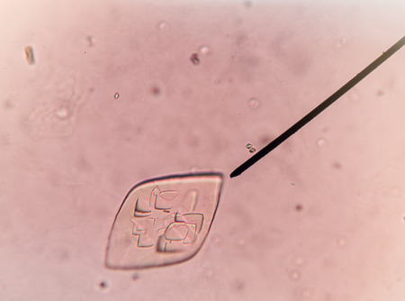
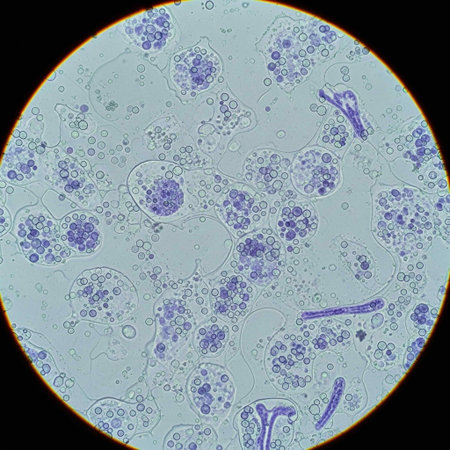
Calcium oxalate in urine find with microscope.
Коллекция по умолчанию
Коллекция по умолчанию
Создать новую
Human cell under microscope.
Коллекция по умолчанию
Коллекция по умолчанию
Создать новую
Digital illustration of white blood cell in color background with blue and purple cells.
Коллекция по умолчанию
Коллекция по умолчанию
Создать новую
Detailed macro image reveals various stages of microscopic embryo development, Ai Generated.
Коллекция по умолчанию
Коллекция по умолчанию
Создать новую
A mesmerizing close-up of colorful bubbles arranged in honeycomb patterns, showcasing intricate details against a vivid pink and black background. Perfect for abstract themes.
Коллекция по умолчанию
Коллекция по умолчанию
Создать новую
Human egg cell, 3D illustration closeup
Коллекция по умолчанию
Коллекция по умолчанию
Создать новую
A stunning microscopic view captures the intricate dance of life as plant cells undergo mitosis. Each stained cell reveals a different stage of division, illustrating the fundamental process of growth and reproduction in a vibrant, detailed display.
Коллекция по умолчанию
Коллекция по умолчанию
Создать новую
Cross-section through the lichen symbiote body 100x
Коллекция по умолчанию
Коллекция по умолчанию
Создать новую
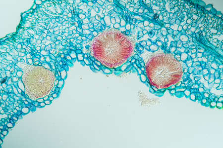
Cowslip stem in cross section 100x
Коллекция по умолчанию
Коллекция по умолчанию
Создать новую
Close-up of pink circular cells under a microscope
Коллекция по умолчанию
Коллекция по умолчанию
Создать новую

micrograph plant tissue, stem of pumpkin
Коллекция по умолчанию
Коллекция по умолчанию
Создать новую

Cross section of human skin under microscope view for education in laboratory.
Коллекция по умолчанию
Коллекция по умолчанию
Создать новую

Field buttercup fruit cross 100x
Коллекция по умолчанию
Коллекция по умолчанию
Создать новую
A detailed, colorful microscopic image of a single cancer cell with numerous tendrils extending outwards, isolated on a clean white background.
Коллекция по умолчанию
Коллекция по умолчанию
Создать новую
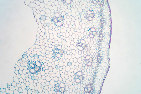
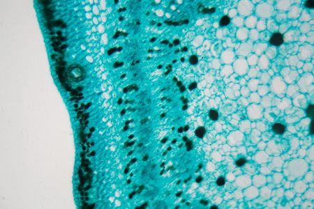
Cross sections of plant stem under microscope view for education plant physiology.
Коллекция по умолчанию
Коллекция по умолчанию
Создать новую
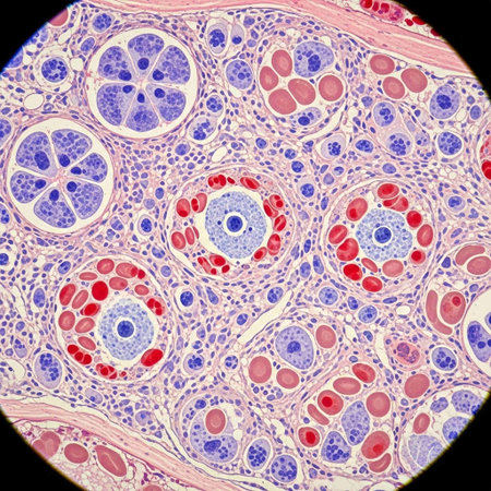
Histopathology of alveoli, light micrograph, photo under microscope
Коллекция по умолчанию
Коллекция по умолчанию
Создать новую
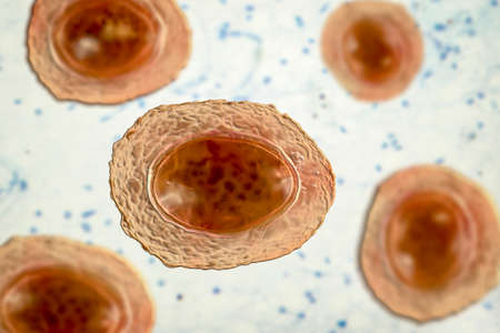
Ascaris lumbricoides, a large roundworm, unfertilized egg, 3D illustration
Коллекция по умолчанию
Коллекция по умолчанию
Создать новую

Cell of human or animal on white background
Коллекция по умолчанию
Коллекция по умолчанию
Создать новую

Cross sections of plant root under microscope view
Коллекция по умолчанию
Коллекция по умолчанию
Создать новую
Bacteria methicillin-resistant Staphylococcus aureus MRSA, multidrug resistant bacteria, 3D illustration
Коллекция по умолчанию
Коллекция по умолчанию
Создать новую
Burdock with stalk across 100x
Коллекция по умолчанию
Коллекция по умолчанию
Создать новую
Rare image of Ghost flatworm - Maricola (Planarian) triclad flatworms in reef aquarium glass
Коллекция по умолчанию
Коллекция по умолчанию
Создать новую
Handmade modern abstract painting. Made with fluid acrylic paints, by acrylic pouring with silicone and torching. Useable as a background or texture.
Коллекция по умолчанию
Коллекция по умолчанию
Создать новую
Cross-sectional margarite flower, 100x
Коллекция по умолчанию
Коллекция по умолчанию
Создать новую
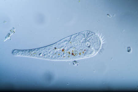
Trumpet animal as a microscopic plankton animal in drops of water
Коллекция по умолчанию
Коллекция по умолчанию
Создать новую
Pancreas cancer cells under microscope view for medical education.
Коллекция по умолчанию
Коллекция по умолчанию
Создать новую
Close up of purple cells under a microscope reveals intricate structures typical of biological lab research.
Коллекция по умолчанию
Коллекция по умолчанию
Создать новую

Cell- science background. Esophagus of the dog- cross section
Коллекция по умолчанию
Коллекция по умолчанию
Создать новую

Host cells with spores (mold) are inside wood under the microscope for education.
Коллекция по умолчанию
Коллекция по умолчанию
Создать новую
Microscopic view showcases colorful cells with unique structures and lively details in biology.
Коллекция по умолчанию
Коллекция по умолчанию
Создать новую
A mesmerizing microscopic view of colorful cells and particles illuminated against a dark backdrop, offering a stunning display of vibrant patterns and structures for scientific exploration.
Коллекция по умолчанию
Коллекция по умолчанию
Создать новую
Microscopy of futuristic genetic engineered cell. Advanced biotechnology concept. Generative AI.
Коллекция по умолчанию
Коллекция по умолчанию
Создать новую
A close up of a cell with many small red and blue dots. The image is a representation of the cell's structure and the different components that make it up
Коллекция по умолчанию
Коллекция по умолчанию
Создать новую
pink organic cells or microorganisms suspended in a fluid, showing biological structures and filaments. Generative AI
Коллекция по умолчанию
Коллекция по умолчанию
Создать новую
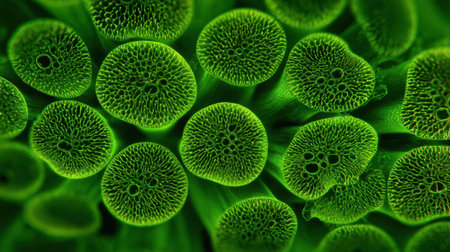
This stunning close-up displays vibrant green algae with intricate round structures. The natural setting emphasizes the beauty of microscopic life, highlighting the details of each cell.
Коллекция по умолчанию
Коллекция по умолчанию
Создать новую
Pancreas cancer cells, light micrograph for medical education.
Коллекция по умолчанию
Коллекция по умолчанию
Создать новую
Microscopic Visualization of Cystopus Candidus Albugo White Rust Fungal Pathogen
Коллекция по умолчанию
Коллекция по умолчанию
Создать новую
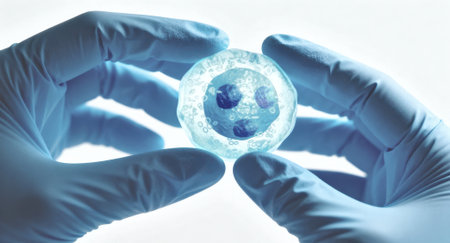
Gloved hands hold a petri dish containing a cultured cell cluster under cool blue backlighting, suggesting laboratory research and biomedical analysis with sterile technique and visible liquid medium ready for examination
Коллекция по умолчанию
Коллекция по умолчанию
Создать новую
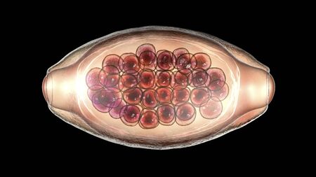
Egg of parasitic roundworm Trichuris trichiura, or whipworm, the causative agent of trichuriasis, disease of a human large intestine, 3D illustration
Коллекция по умолчанию
Коллекция по умолчанию
Создать новую
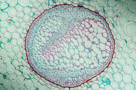
Fern stems in cross section 100x
Коллекция по умолчанию
Коллекция по умолчанию
Создать новую
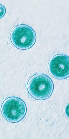
Microscopic view of teal round parasitic oocysts with visible internal cellular structures against a textured light-blue background, science and microbiology research imagery
Коллекция по умолчанию
Коллекция по умолчанию
Создать новую
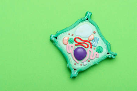
Model of plant cell on green background. Green world and education concept
Коллекция по умолчанию
Коллекция по умолчанию
Создать новую
Structure of human cell under microscope view. 3D illustration.
Коллекция по умолчанию
Коллекция по умолчанию
Создать новую
Johannes berry fruit cross 100x
Коллекция по умолчанию
Коллекция по умолчанию
Создать новую
Microscope photo of a cross section of a cotton stem.
Коллекция по умолчанию
Коллекция по умолчанию
Создать новую
a close up of a colorful plant
Коллекция по умолчанию
Коллекция по умолчанию
Создать новую
A microscope slide containing a sample of plankton viewed under high magnification to study its composition
Коллекция по умолчанию
Коллекция по умолчанию
Создать новую
An intricate view of the insides of an embryo or bacteria contained within the cell wall, through a microscope. Stylized and colourized interpretation.
Коллекция по умолчанию
Коллекция по умолчанию
Создать новую
Intricate world of living cells in this closeup, showcasing glowing organic structures on a stunning blue background, revealing lives microscopic beauty
Коллекция по умолчанию
Коллекция по умолчанию
Создать новую
Legion-Media
Создайте свои проекты на основе качественных стоковых фотографий и видео.


Copyright © Legion-Media.